
Multimodality Cardiovascular Imaging

Our research group develops and applies non-invasive cardiovascular imaging to characterize the mechanisms, progression and clinical consequences of cardiovascular disease. We combine cardiac magnetic resonance imaging (CMR), cardiac computed tomography and echocardiography with clinical information, biomarkers and longitudinal outcomes. Our aim is to identify imaging markers that explain symptoms, detect early disease and improve individual risk assessment. The clinical focus includes hypertrophic cardiomyopathy (HCM), heart failure with preserved ejection fraction (HFpEF), atrial fibrillation and atrial cardiomyopathy, as well as structural and valvular heart disease.
We also investigate the interaction between atrial and ventricular function, the pulmonary circulation and systemic vascular loading. Our work examines atrial deformation and three-dimensional remodeling, right ventricular reserve and ventricular–vascular coupling.
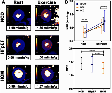
These measurements help characterize disease that may be difficult to recognize using conventional resting parameters. In HCM, multicenter clinical cohorts complement functional imaging by relating disease phenotypes to subsequent heart failure and arrhythmic outcomes.
Quantitative tissue characterization is another major focus. We study myocardial fibrosis, perfusion and epicardial adipose tissue to understand how tissue composition relates to cardiac remodeling and function. Epicardial adipose tissue is of particular interest because of its close anatomical relationship with the myocardium. Our projects address reproducible volumetric quantification using CMR and CT, its association with functional impairment, and its potential diagnostic and prognostic relevance in heart failure and aortic valve disease.
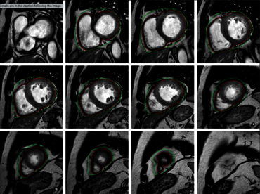
To make quantitative imaging more reproducible and applicable to larger cohorts, we combine automated image analysis, artificial intelligence and modern statistical methods. Current projects include image segmentation, radiomics and data-driven phenotyping. We place particular emphasis on image quality, methodological validation and testing against clinically meaningful outcomes. This approach connects technical development with the requirements of clinical cardiovascular research.
Our long-term goal is to translate validated imaging methods and quantitative biomarkers into clinical practice. Through interdisciplinary and multicenter collaboration, we aim to support earlier diagnosis, more precise risk stratification and treatment decisions informed by the predominant disease mechanism. Within the HBCG, this work provides a cardiovascular imaging framework for collaborative studies of cardiac remodeling and its wider clinical consequences.
Selected recent publications
- Midventricular Obstruction in Hypertrophic Cardiomyopathy: Clinical Outcomes in a Multicenter Cohort.
Schulz A, Rowin EJ, Peng LW, et al.
Journal of the American College of Cardiology. 2026.
This multicenter study characterizes the clinical course of HCM with midventricular obstruction, highlighting the importance of recognizing this distinct phenotype when assessing heart failure progression and arrhythmic risk.
- Quantitative myocardial blood flow and perfusion reserve with exercise cardiovascular magnetic resonance.
Schulz A, Wallace TE, Chow K, et al.
Journal of Cardiovascular Magnetic Resonance. 2025;27(2):101988.
This pilot study demonstrated the feasibility of quantitative post-exercise perfusion CMR after image quality control. Patients with HCM showed lower post-exercise myocardial blood flow and perfusion reserve than patients with HFpEF or non-cardiac dyspnea.
- Left Atrial Roof Enlargement Is a Distinct Feature of Heart Failure With Preserved Ejection Fraction.
Backhaus SJ, Nasopoulou A, Lange T, Schulz A, et al.
Circulation: Cardiovascular Imaging. 2024;17(7):e016424.
Three-dimensional CMR models identified atrial roof dilation in patients whose HFpEF was diagnosed only during exercise. The derived atrial shape score was associated with atrial fibrillation and cardiovascular hospitalization.
- Concomitant latent pulmonary vascular disease leads to impaired global cardiac performance in heart failure with preserved ejection fraction.
Schuster A, Schulz A, Lange T, et al.
European Journal of Heart Failure. 2023;25(3):322–331.
Latent pulmonary vascular disease was associated with impaired right ventricular reserve, reduced left atrial filling and lower cardiac output. Exercise CMR characterized these abnormalities, illustrating the contribution of the pulmonary circulation to functional limitation in HFpEF.
- RT-CMR Imaging for Noninvasive Characterization of HFpEF: Medium-Term Outcomes of the HFpEF Stress Trial.
Backhaus SJ, Rösel SF, Schulz A, et al.
JACC: Cardiovascular Imaging. 2022;15(5):943–945.
The medium-term follow-up of the HFpEF Stress Trial demonstrated the prognostic relevance of CMR-derived atrial strain, connecting detailed functional characterization with subsequent clinical outcomes.
Team

contact information
- telephone: +49 551 3966315
- e-mail address: alexander.schulz(at)med.uni-goettingen.de